


With serious internal diseases, poor nutrition, as well as with age, the growth of the nail slows down, its structure changes. Only a doctor can determine the exact cause of the violation based on the test results and microscopic examination.
But to know what will happen to the nails in the legs or hands, you can use photos of different types of fungal diseases.
Causes of nail deformation
Molds, yeast-like fungi and dermatophyte that cause infectious nail diseases (onychomycosis), have similar symptoms.
All toenail fungus or nail fungus that deform the nail, change its transparency, shine, and color, can be seen in the photos presented.
Nail changes occur not only with onychomycosis but also with trauma, chronic mental illness (nail fold inflammation), psoriasis, eczema, dermatitis. Before concluding that you have a fungal infection, you need to consider all possible options.
Signs of fungal infection
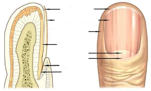
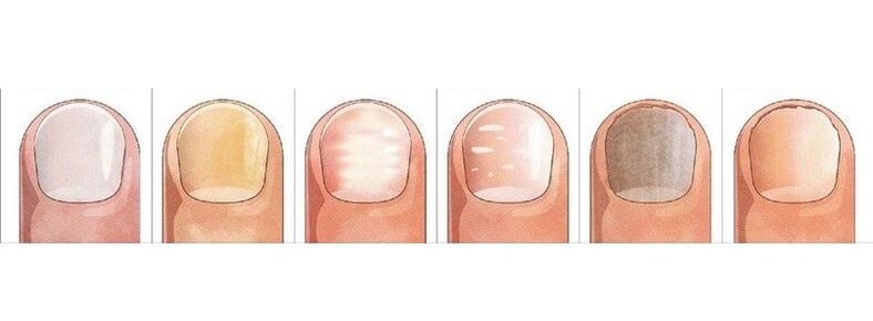
The most informative signs of a fungal infection are nail color change, nail peeling, change in appearance - transverse grooves, longitudinal grooves, dents, thickening, destruction of the nail.
The pink color of a healthy nail is determined by the transparency of the nail and the visible blood vessels through it. With onychomycosis, the nail loses its transparency, the color becomes brown, yellow, rarely green or black.
Candida fungi and dermatophytes cause onychomycosis - separating the affected part of the nail. With fungal skin infections, onychomycosis can be observed from the far edge of the nail, and when infected with Candida, the nail lags behind the nail bed at the base, in the crescent-shaped area.
A symptom of candidiasis can be inflammation of the paronychia. It has bacterial forms caused by streptococci and staphylococci, as well as non-infectious forms - eczema, psoriasis, systemic vasculitis.
When the toenail is affected by Trichophyton rubrum, the sheet is affected, as you can see in the photo, the roller is not affected by the infection. The plaques become yellowish, strongly thickened, the accumulating fungal masses are clearly distinguished below it.
Nail fungus caused by a dermatophyte infection
In 95% of all nail fungus cases, the disease is caused by the fungus Dermatophytes Trichophyton rubrum and Trichophyton mentagrophytes.
Trichophyton whip infection
Onychomycosis begins when a fungus enters underneath the nail plate from the free margin. Fungal infection is manifested by the appearance of a yellow spot, uneven, crumbling surface at the far (distal) edge of the nail in the speckled area.
far-side formatof Trichophyton rubrum dermatological fungal infection is common. In the photo, you can see the fungal infestation marks along the folds around the lateral nail.
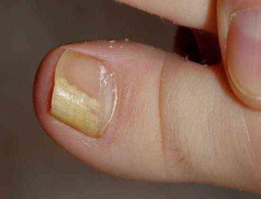
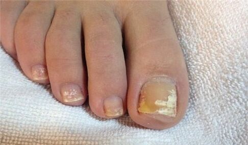
As a rule, Trichophyton rubrum affects the big toe, causing hyperkeratosis - a fungal buildup between the nail and the nail layer, which looks like a yellowish liquid in the photo.
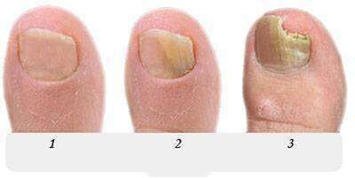
At this stage, the fungus occupies a negligible part of the nail, as in the photo shown, and with the help of topical treatment, it is possible to cope with onychomycosis initially.
If left untreated, the stain will grow, gradually affecting the entire edge of the nail, then turning into a semicircle. In the photo, the onychomycosis looks like yellow stripes towards the growing area of the nail plate.
Withdistal form of onychomycosis, commonly found on the big toe, a yellow infectious spot appears on the far edge of the nail, in its central part, possiblysee in photo.
In the severe stage of the fungus of the legs, some nails are affected, as shown in the photo, and the treatment is no longer limited to topical medications and medications. In addition to the antifungal, the nail is hard-cleaned, to remove all or part of the nail.
Long-term therapy with the use of all known drugs and antifungal treatments will be performed on the legs, caused by Trichophyton rubrum, with hyperkeratosis, as can be seen in the photo.
Fungal infection with total nail damage spreads to the entire nail area, the nail is completely destroyed.
Infection with another representative of the fungus dermatophytes, the fungus Trichophyton mentagrophytes, can also lead to a whole nail fungal infection.
Infections Trichophyton mentagrophytes
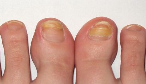
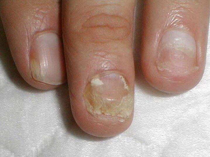
With the toenails completely defeated by Trichophyton mentagrophytes, deformed nails, photos showed that the nails thickened, changed structure, flattened, the entire nail surface appeared yellow spots.
This nail infection usually causes white, superficial fungal disease on the big toe, less common in the little toe.
This fungus does not actually occur on the nails, it often causes fungal skin diseases of the feet, as shown in the photo, and requires treatment of the skin on the feet and nails at the same time.
A symptom of a fungal nail infection, often on the feet, are white spots of various sizes, as shown in the photo, reminiscent of leukemia - a disease of the nail itself.
But unlike leukocytes, in which white spots are caused by the appearance of air bubbles in the layers of the nail, white spots in fungal infections are the result of Trichophyton's actionmentagrophytes.
Rarely, superficial white onychomycosis is caused by mildew; In AIDS, the causative agent of this fungus can be Trichophyton rubrum and affects the nails in both feet and hands.
Nail changes due to Candida infection
The fungus is more common in women, and it affects the nails on working hands that are frequently exposed to water.
For onychomycosis, a proximal form of infection is characteristic, in which the fungus affects the folds of the nail first at the base of the nail, then penetrates the growing area and the nail bed. It then gradually moves along the foundation from base to edge, occupying an ever larger area of the foundation plate.
The causative agent of onychomycosis is the fungus Candida albicans. This fungus penetrates the toenails and nails, spreading from the semi-circle at the base of the nail, to the free edge, as you can see in the photo.
Signs of onychomycosisalbicans is inflammation of the nail folds (paronychia), separating the epidermis from the nail, pain, discharge when infected with bacteria. .
Candida albicans can penetrate the nail and from its free margin. In this case, they talk about the distal form of infection, which is associated, as a rule, with candidiasis of the skin.
Treating candidiasis on nails and feet with damage to more than half of the area of the nail plate, as shown in the photo, not only includes the fight against onychomycosis, but also measures to reduceCandidiasis activity in their natural reserves - intestine, oral cavity, genital mucosa. . .
Fungal infections
Mold causes fungus less often than Candida or ringworm. The main symptom of a fungal toenail infection, as you can see in the photo, whenchanges the nail color to blue, black, and green.
Signs of toenail fungus can be black spots, a dot on the nail plate or as shown in the photo a black vertical stripe.
Antifungal preparations
Antifungal pills with fluconazole, ketoconazole, terbinafine, itraconazole, griseofulvin are used to treat nail fungus caused by dermatophytes, such as in this photo.
Antifungals with terbinafine are effective against dermatological infections.
Antifungal drug with voriconazole is highly active against dermatophytes.
Itis used andto treat nail moldon feet, hands andagainst candida yeast. The effect spectrum includes molds such as Aspergillum, Fusarium, Penicillium.
Itraconazole-based preparations for dealing with mold.
Fungal nail diseases
Occasionally appears grayish coloronnails with eczema. In this case, the nail can move away from the nail layer, where fungus can be observed.
The appearance is very similar to the onychomycosisof psoriasis. With this disease, not only doeschange colorbutnails also thicken.
Concave points are found on its surface, the separation of the foundation plate from the foundation is noted. But there is a difference from fungi: in psoriasis, separate and healed toenails are separated by a yellow, pink band over time.
Bluemanicurefor fake nail infection. Regular mechanical friction against the nail folds causes the appearance of surface grooves, roughness of the nail.
White spots of leukocytes, appearance ofis associated with metabolic disorders, can also be mistaken for a superficial white fungus with a large spot area.
Changes the color or shape of the nail causing injury. The big toe is most at risk. The nail is traumatized as well as fungal, thickening and darkening.

The difference between a wound and a fungus is that changes in the wound process are noted only on the injured finger, the nails on the other fingers remain unchanged, and there is no infection from the fingerdisease as in onychomycosis.
The result of injury can be a partial separation of the nail from the nail layer, forming a cavity, under unfavorable conditions, the fungus will quickly colonize.
The nail plate can be separated from the nail layer under the action of light (photolysis), iron deficiency anemia, hormonal diseases. Separation and loss of nails occur with lichen lichen, bullous skin, trauma to the nail.
But in the end you can be sure that the conclusion is correct and starting treatment, you can only make it after seeking help from a dermatologist - a specialist in the diseaseskin or fungal doctor - doctors who treat fungal diseases.